apical lung hernia
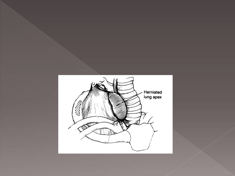
Pulmonary herniation is an extension of the lung and pleura beyond their native positions in the thoracic cavity. 1 A hernia involving the cervical space has also been described as apical in the literature.

Right Upper Lobe Pneumonia Pneumonia Radiology Medicine
Airway fluoroscopy or CT pe.
. Hernia of the lung occurs infrequently and not all of those that do occur cause symptoms that require treatment. Symptoms when reported tend to be due to extrinsic pressure from the hernia on neck structures eg. Radiologic studies performed at midinspiration may not show hernias.
Apical lung herniation in adults is rare particularly in the absence of penetrating lung injury or chest wall disease. Sometimes the diagnosis can only be made with a Valsalva maneuver which accentuates the herniation improving its visibility on physical examination. Apical lung hernias typically manifest as unilateral right-sided air radi- olucencies at the thoracic inlet on chest radiographs.
View this article on Wiley Online Library. Request PDF On Oct 1 2007 Jyotsna M Joshi published Apical lung hernia. Symptoms when reported tend to be due to extrinsic pressure from the hernia on neck structures eg.
Article in Italian Authors F Zamparelli 1 G Turtulici T Luminati E Tagliafico C Frola. 1Respiratory Medicine BYL Nair Hospital and Topiwala National Medical College Mumbai Maharashtra India. Download Citation On Aug 1 2008 George R Crowe published Re.
Apical lung herniation Thorax. Apical lung herniation in children is perhaps due to disproportionate growth of the ribcage and often presents early then gradually resolves with the babys growth. In rare instances a lung hernia may become strangulated.
This lung herniation was probably caused by a congenital deficiency in the suprapleural membrane Sibsons fascia combined with increased thoracic pressure created by the respiratory tract infection. Apical lung hernia Find read and cite all the research you need on ResearchGate. Dysphagia esophageal or coughing trachea 2.
Herniae are defined as a herniation of the lung beyond the confines of the thoracic cage. They are frequently intermittent and can cause lateral tracheal deviation. Find read and cite all the research you need on ResearchGate.
Lung herniation by itself remains asymptomatic unless complicated by secondary factors like external injury or compression upon neck structures. 1 2 it is due to a defect in the suprapleural membrane sibsons fascia and small incidental apical parietal pleural defects have been described which may be present prior to the development of a larger defect. About half of all lung hernias however appear after trauma to the chest 5.
This is a rare pathological entity with fewer than 300 reported cases in the literature presented mainly as a single case reports12 According to their location lung hernias can be cervical intercostal or diaphragmatic and each of these types is. Apical lung hernia is a rare variety and has been confined to few case reports and series. The term pathological hernia is used to describe lung herniation induced by conditions such as tuberculosis focal bacterial infections empyema or osteomyelitis and malignant diseases 1.
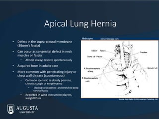
Herniation occurs through a defect in the Sibsons fascia and the apical segment of the lung protrudes in between the scalenus anterior and sternocleidomastoid muscles. Apical lung hernias typically manifest as unilateral right-sided air radiolucencies at the thoracic inlet on chest radiographs. Pulmonary hernias have been described through the diaphragm intercostal spaces and into the cervical space.
Sometimes the diagnosis can only be made with a Valsalva manoeuvre which accentuates the herniation improving its visibility on physical examination. Apical lung hernias are often asymptomatic 1-3. Surgical repair was not necessary as the hernias were asymptomatic and not associated with chronic cough.
17874988 Indexed for MEDLINE Publication Types. Affiliation 1 Ospedale Evangelico. Radiologic studies performed at midinspiration may not show hernias.
3 4 tearing of. Authors Harpreet Ranu 1 Mark Jackson. Lung herniation is defined as a protrusion of the lung beyond the normal confines of the thoracic cavity.
Dysphagia oesophageal or coughing trachea 2. Spontaneous apical lung herniation presenting as a neck lump in a patient with Ehlers-Danlos syndrome Ehlers-Danlos syndrome EDS is a heritable group of disorders of connective tissue characterised by skin hyperlaxity joint hypermobility and tissue fragility. Apical lung hernias typically manifest as unilateral right-sided air radiolucencies at the thoracic inlet on chest radiographs.
Description of a case studied with spiral computerized tomography and tridimensional reconstruction Radiol Med. They are frequently intermittent and can cause lateral tracheal deviation. They are frequently intermittent and.
PDF On Feb 10 2014 Srikanth Prasad and others published An unusual cause for neck swelling. Epub 2011 Apr 20. Lung hernia Last revised by Dr Yuranga Weerakkody on 24 Oct 2021 Edit article Citation DOI article data Lung hernias alternative plural.
Apical lung hernia. Apical lung hernia Find read and cite all the research you need on ResearchGate. They are uncommon mostly seen post trauma or thoracotomies.
Affiliation 1 Department of Respiratory. Some hernias of the lung however are symptomatic being accompanied by local pain paroxysmal coughing hemoptysis or any combination of the three. Apical lung hernias are often asymptomatic 1-3.

Bilateral Cervical Lung Hernia With T1 Nerve Compression The Annals Of Thoracic Surgery

Is Collapse Of The Lung With Increased Lucency On A Chest X Ray Always A Pneumothorax Journal Of Cardiothoracic And Vascular Anesthesia

Pdf Dynamic Cervical Lung Lobe Herniation In A Shih Tzu Dog Semantic Scholar

Pdf Adult Cervical Lung Herniation Importance Of Valsalva Manoeuvre In Imaging Semantic Scholar

Right Sided Congestive Heart Failure Secondary To Supraventricular Tachycardia In A Dog With A Right Atrial Mass Basili 2021 Veterinary Record Case Reports Wiley Online Library
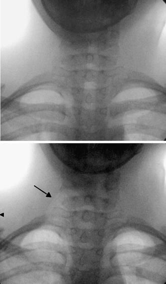
Dynamic Cervical Lung Herniation In A 10 Year Old Girl With Cough Springerlink

Chest X Ray Showed Apical Mass On The Right Lung No Pleural Effusion Download Scientific Diagram

Extra Luminal Contrast Leakage Was Detected On The Initial Esophagogram Download Scientific Diagram
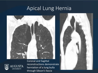
Interesting Cases Of Lung Hernias

Apical Lung Herniation Radiology Case Radiopaedia Org
Article Medicale Tunisie Article Medicale

Interesting Cases Of Lung Hernias

Pdf Dynamic Cervical Lung Lobe Herniation In A Shih Tzu Dog Semantic Scholar

45 Year Old Asymptomatic Man With Right Apical Lung Hernia Download Scientific Diagram

Imaging Of The Neonate Term Infant Sciencedirect
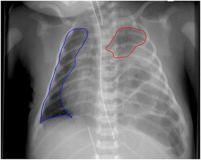
Frontiers The Chest Radiographic Thoracic Area Can Serve As A Prediction Marker For Morbidity And Mortality In Infants With Congenital Diaphragmatic Hernia

Intercostal Lung Hernia Radiographic And Mdct Findings Clinical Radiology
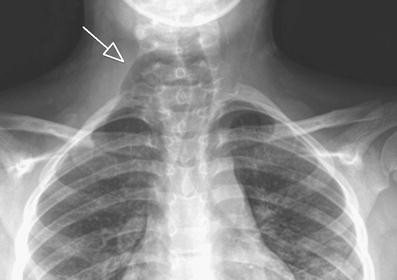
Dynamic Cervical Lung Herniation In A 10 Year Old Girl With Cough Springerlink
Comments
Post a Comment